
Researching FTD: Brain Scans and Blood Tests
Researchers use different tools to understand frontotemporal dementia (FTD), including brain scans and blood tests. MRI and PET scans provide complementary information: MRI shows the brain’s structure, while PET can reveal changes in processes such as energy use or inflammation. Blood tests can help researchers investigate genetic causes, proteins linked to disease processes and immune responses.
MRI: Looking at Brain Structure
Researchers want to understand what is happening in the brain across the course of FTD, including before and after symptoms begin. Brain-imaging techniques allow us to do this without surgery or other invasive procedures.
Magnetic Resonance Imaging, or MRI, is used routinely in clinical care and research to produce detailed images of structures inside the body. While X-rays are particularly useful for looking at bones, MRI is especially good at imaging soft tissues, including the brain.
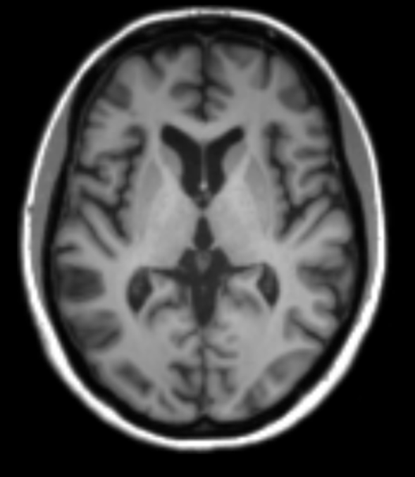
An MRI scanner uses a powerful magnet and radio waves to create detailed three-dimensional images. In clinical care, these images can support diagnosis by identifying patterns of brain change that may be associated with FTD. For example, some forms of FTD are linked to shrinkage in particular areas of the frontal and temporal lobes, which are involved in behaviour, language, planning and social understanding.
In research, MRI allows us to measure these changes precisely and to investigate how they relate to symptoms, biomarkers and disease progression. Researchers can use specialist MRI techniques to assess:
- Brain volume and atrophy, meaning loss of brain tissue over time
- The structure and connectivity of brain regions
- Blood flow and blood vessels within the brain
- Brain activity or function in particular regions
MRI does not use ionising radiation. However, because it uses a strong magnet, it is important to remove metal items such as jewellery, watches, hearing aids and clothing with metal components before the scan. If you have an implant, such as a pacemaker, cochlear implant, metal clip or joint replacement, the research team will check whether it is safe for you to have an MRI. Eligibility is assessed individually.
During the scan, you will lie on a padded bed that moves slowly into the scanner. For brain imaging, the radiographer will place a device called a head coil around your head. Despite its name, it does not touch or squeeze the head, it helps the scanner collect clearer images.
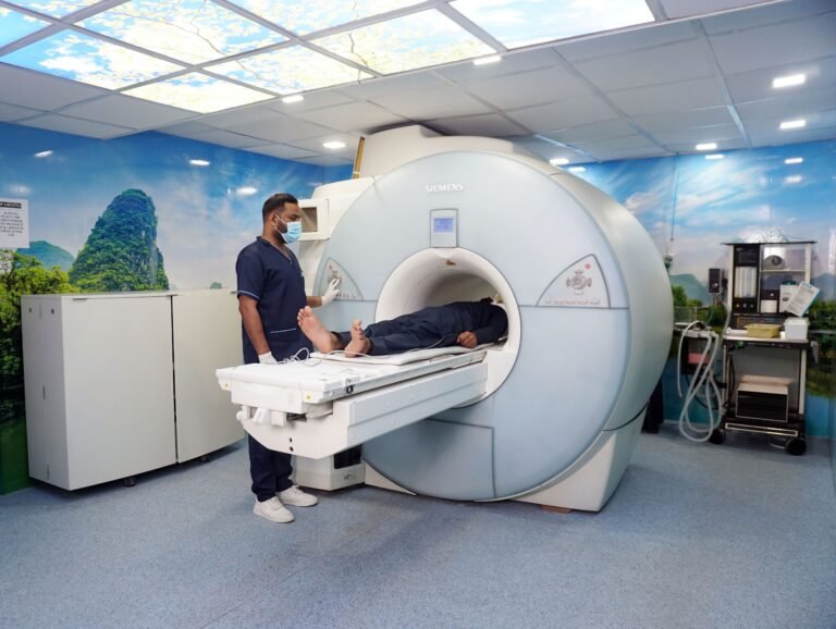
It is important to remain as still as possible, as movement can reduce image quality. The scanner can be noisy, producing loud tapping or knocking sounds while images are being collected. Centres may provide earplugs, headphones or music to make the experience more comfortable. You can speak with the radiographer throughout the scan, and you will be given a call button or buzzer so that you can stop the scan or ask for assistance at any point.
PET: Looking at Brain Biology
MRI cannot show every biological process that researchers want to study. Positron Emission Tomography, or PET, is an imaging technique that can help researchers examine specific processes occurring in the brain.
PET involves a small injection containing a radiotracer. A radiotracer is a substance designed to bind to, or be taken up by, a particular biological target. The scanner detects the tracer and creates an image showing where it has travelled in the body or brain. Different tracers can be used to study different aspects of brain biology.
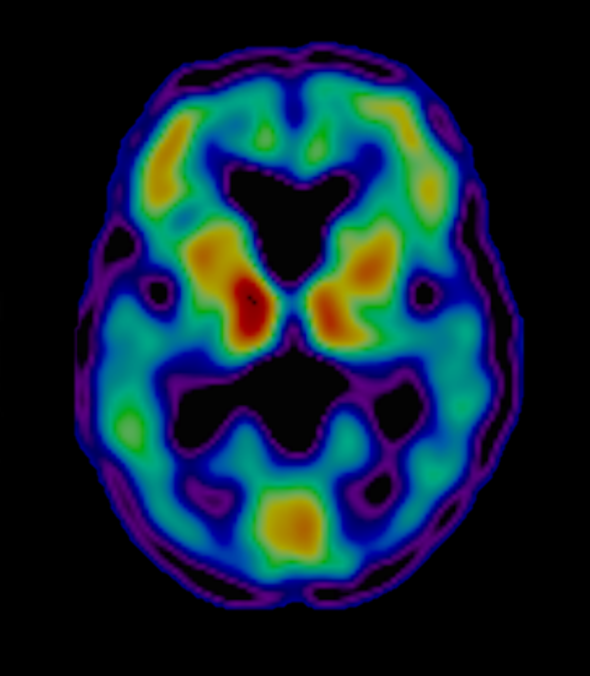
One PET scan commonly used in FTD research is FDG-PET. FDG is a form of glucose labelled with a small amount of radioactivity. Because the brain needs glucose for energy, FDG-PET provides a measure of how much glucose different brain regions are using.
Areas affected by FTD may use less glucose than expected. This appears on an FDG-PET scan as regions of reduced signal, sometimes described as reduced metabolism. These changes may be seen in frontal or temporal parts of the brain, depending on the type of FTD. In some people, altered brain metabolism can be detected before clear loss of brain tissue is visible on MRI.
At ON-FIRE, we are particularly interested in inflammation in the brain and its potential role in FTD. Using specialist PET tracers, researchers can study biological signals linked to inflammatory processes. This may help us understand where inflammation is occurring, how it changes over time, and whether it is related to symptoms, protein changes, brain atrophy or other features of FTD.
Why combine MRI and PET?
MRI and PET provide different, complementary information. MRI gives a detailed map of the brain’s structure, while PET can show changes in metabolism or other biological processes.
Researchers often combine these methods to build a more complete picture. For example, an MRI scan can help us identify the exact brain location of a PET signal and account for any tissue loss in that area. Together, the scans may help researchers distinguish between changes in brain structure, brain function and underlying biology.
MRI (left) and TSPO-PET (right) brain scans. Slide to move between the images.
What happens during a PET scan?
Before a PET scan, the research team will review whether the scan and tracer are suitable for you. This includes questions about general health, medications and, where relevant, pregnancy or breastfeeding.
The exact procedure depends on the tracer being used, but it usually involves two stages:
- Tracer injection and uptake period. For some PET scans, the radiotracer is injected first and there is a waiting period while it circulates and reaches its target in the brain. This is known as the uptake period.
- The scan itself. You will then lie still on a scanning bed while images are acquired. For other tracers, the injection may take place while you are already in the scanner.
The radiation dose from a research PET scan depends on the specific tracer and scan protocol. It is carefully calculated and reviewed by the research and clinical teams, and approved by committees. The tracer naturally decays and leaves the body over time; for most PET tracers used in research, very little remains after about 24 hours. The study team will give participants clear, tracer-specific instructions about any precautions after the scan.
By studying brain structure alongside processes such as metabolism and inflammation, researchers hope to identify new biomarkers, tools for better diagnosis and treatment targets for FTD.
Blood
Blood tests are familiar, relatively quick and widely used in healthcare. Researchers can also use blood samples to study biological changes that may be associated with FTD. At ON-FIRE, we are working to discover and evaluate blood-based markers that could help us understand FTD and, in time, support better diagnosis or monitoring.
A blood sample can be analysed in different ways. In this section, we focus on three examples: genetic testing, protein and other molecular markers, and immune-cell analysis.
What happens during a blood test?
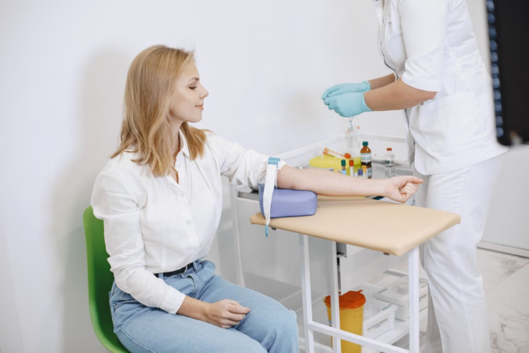
A trained member of the research or clinical team takes a small sample of blood, usually from a vein in your arm. The needle may cause a brief sharp scratch, and some people have temporary bruising or soreness afterwards. Several tubes may be collected so researchers can carry out different tests, or store samples for future analyses, if the study consent allows this.
Genetic Testing
FTD can have a genetic cause, but not everyone with FTD has an identifiable disease-causing genetic change. Estimates vary between studies and populations. Around one in five people with FTD may have an underlying genetic cause. The genes most often associated with inherited FTD are C9orf72, GRN and MAPT.
Genetic testing can help identify whether a person’s FTD is linked to a known genetic change. Testing may be considered based on a person’s symptoms and family history, but family history alone does not determine whether testing is appropriate. A genetic specialist can explain what a test may, and may not, tell someone.
A result can also have implications for biological relatives. Some people want to know whether they carry a genetic change, others would rather not know. Genetic counselling gives people the opportunity to discuss these issues and consider their options before deciding whether to be tested.
For many genetic tests, a blood sample is used to obtain DNA. The laboratory then examines relevant genes for changes known to be associated with FTD. The approach depends on the gene. For example, some C9orf72 tests look for an unusually long repeated section of DNA.
Proteins and other substances
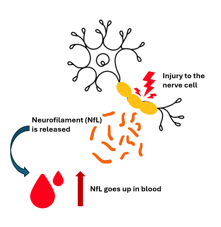
Researchers can measure proteins and other substances in blood that may reflect processes occurring in the brain or elsewhere in the body. One example is neurofilament light chain, often shortened to NfL. NfL is a protein found inside nerve cells, and when these cells are injured, NfL can enter the blood. Higher levels can therefore indicate nerve-cell damage.
NfL is being studied as a marker of neurodegeneration and may help researchers track disease-related change. However, it is not specific to FTD: levels can rise in different neurological conditions and for other reasons. On its own, an NfL result cannot diagnose FTD or reliably identify the cause of someone’s symptoms.
Some blood markers are more closely linked to particular diseases. For example, blood tests measuring forms of phosphorylated tau, including p-tau217, are being used in some clinical settings to help assess the likelihood of Alzheimer’s-related changes. Their use depends on the specific test and clinical context.
ON-FIRE is investigating whether additional blood-based markers could help researchers study FTD across different genetic forms and in people whose condition has no known genetic cause. Researchers are also interested in whether blood markers might reveal more about disease processes – not just help with diagnosis.
Immune cells and proteins
At ON-FIRE, we are also interested in inflammation and the immune system. Researchers can study immune-related proteins in blood, or examine immune cells directly.
Different types of immune cells have different roles and work together to protect the body from infection, injury and other threats. One way to picture this is as an orchestra: each cell type is like a different instrument, contributing to the overall performance. If one part changes, the balance may shift. In a similar way, changes in the number or behaviour of immune cells may provide clues about what is happening in the body, although they do not by themselves show that inflammation is causing FTD.

Researchers can compare immune cells and inflammation-related proteins in people with FTD and people without the condition, and between people with different disease trajectories. If a difference is found, further studies are needed to understand what it means and whether it is related to FTD, another health factor, or both. Over time, this work may help identify biological processes that could be explored as potential treatment targets.
Why blood tests matter
Genetic testing, protein measurements and immune-cell analysis each offer a different view of FTD. Together, they may help researchers study inherited risk, changes associated with nerve-cell injury, and the body’s immune response.
Blood tests are generally easier to collect than PET scans and may be more practical to use at scale. But a promising research result is not automatically ready for routine healthcare: each test needs to be carefully validated to show what it can reliably tell us, for whom, and how it should be used. The long-term aim is to develop accessible tools that complement clinical assessment and imaging – not replace them – and help improve understanding of FTD.